Project supported by AMI could make it easier to diagnose UTIs
A project supported by Applied Microbiology International’s Small Research Projects and Equipment grant shows promise in identifying microbe-borne biomarkers for urinary tract infections.
We highly appreciate the AMI Small Research Projects and Equipment grant as it enabled us to combine microbiological, immunological and proteomics analysis to advance the scientific impact of the study on the marker of urinary tract infection.
The objective of the project was to characterise proteins of bacterial biofilm specific for urinary tract infections (UTIs) and to select useful microbe-borne biomarkers. The funds were used to purchase consumables crucial for proteomic analysis.
Seeking accuracy
Urinary tract infections are amongst the most common infections worldwide. Till now, urine culture is considered as the gold standard for the diagnosis of UTI but their diagnostic accuracy can be lower in specific groups of patients, such as infants or patients with continuous catheterization, immunosuppression or RTx recipients.
As an alternative, the authors of the current project have focused on the monocytes activation to estimate the urine isolates’ pathogenicity. The results showed promising predictive value of the technique in the diagnosis of urinary tract infection. It was hypothesized then, that the activation of monocytes results from the synthesis of particular proteins within the biofilm.
The above assumption was supported by the fact that thousands of particular proteins can be identified in urine using shotgun proteomics which makes the identification of such microbe-borne UTI biomarkers very likely. Furthermore, the direct detection of such proteins could make the UTI diagnosis simpler, and faster. Over and above it could scientifically contribute to the development of innovative diagnostic procedures also.
Urine samples
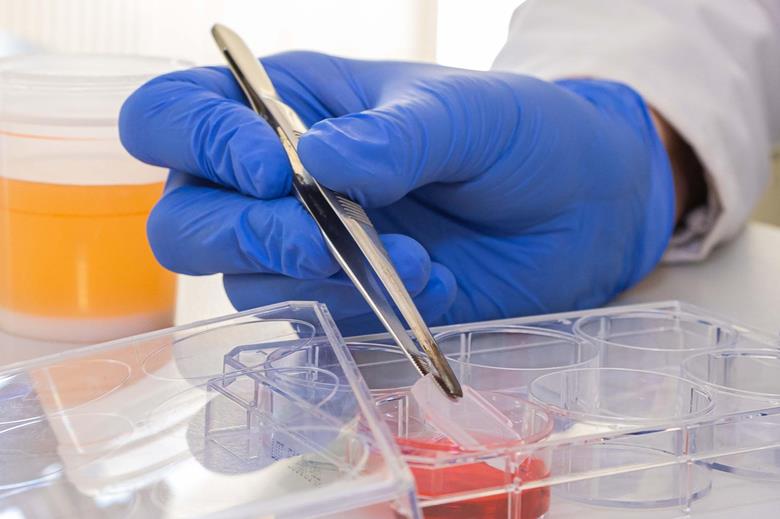
Microbiologically negative (10) and positive urine samples (CFU/ml>10*6 E. coli x 10; CFU/ml>10*6 E. faecalis x 10 were collected from the patients of University Clinical Centre, Medical University of Gdansk.
In the first step, the urine collected in the mid-stream procedure was inoculated on the 6-well plate and then incubated for 72h to let bacteria form the biofilm. Then the urine was removed and stored for the proteomic analysis and the plate with biofilm was used for microbe-immune interactions analysis.
For proteomic analysis, urine samples were supplemented with boric acid, centrifuged and filtered through a dedicated membrane. The next steps of proteomic analysis were progressed by the researchers of Department of Chemistry at Nova School of Science and Technology | FCT NOVA Universidade - Hugo M. Santos and José Luis Capelo Martínez.
Treating biofilm samples
The biofilm sample and bacterial pellet were lysed using reducing SDS Tris-HCl buffer. After the addition of the reducing Lysis Buffer, samples were incubated at 95ºC, followed by sonication. Then, we performed a Filter-Aided Sample Preparation (FASP) proteome digestion.
Samples were analysed by LC-MS/MS. All MS/MS spectra were exported into Mascot Generic Files (.mgf) files for data analysis by applying an internal script from Data Analysis 4.2 (Bruker). Data analysis was performed using Mascot 2.3.02 (Matrix Science, London, UK) integrated into Protein Scape 4.0 (Bruker).
Search parameters were as follows: UniprotSwissProt FASTA file, trypsin/P was selected as the digestion enzyme, and two missed-cleavages were allowed. Monoisotopic peptide masses were searched with 25 ppm peptide mass tolerance and 0.05 Da fragment mass tolerance. Carbamidomethyl on Cysteine was selected as a fixed modification, and oxidation on Methionine and Acetylation of protein N-terminal as the variable modifications.
Microbe-immune interactions
Simultaneously, to analyse microbe-immune interactions we washed out the plate with biofilm with the saline and then the plate was filled with the suspension of human monocytes The plate was then incubated for 60 minutes and the suspension of monocytes was aspirated for flow cytometry analysis.
Monocytes’ response to the bacterial biofilm was estimated as the rate of monocytes’ adhesion to biofilm (measured as a difference between inoculum of culture before and after 60 minutes incubation), changes of monocytes’ size (by using FSC flow cytometry parameter) and the cytotoxicity of the biofilm.
The last parameter was estimated as the change of monocytes’ cell membrane permeability and measured as the increase of propidium iodide fluorescence intensity). After the flow cytometry analysis, the biofilm was removed from the plate and lyophilized before shipment to Portugal for proteomics analysis.
Proteomic analysis
Proteomic analysis of the samples resulted in the detection and identification of 118 microbial proteins including protein known to be related to pathogenicity of the strains and biofilm formation as elongation factor Tu, division protein divIVA and mechanosensitive channels. Coincidence of virulence estimated by monocyte activation and deduction of specific proteins makes the result promising.
Further analysis of collected data and evaluation of proteins not categorized yet will hopefully result in selection of the unique markers of urinary tract infection.
Last but not least, the funds provided by AMI allowed us to set meaningful and essential collaboration between the study group from the Medical University of Gdańsk and the leading experts of proteomic analysis - BIOSCOPE Research Group from University Nova of Lisbon, Portugal.
Agnieszka Daca, student
Tomasz Jarzembowski, supervisor